Home » Without Label » Bones In Leg Diagram / Broken Leg Pictures Causes Treatment Surgery Recovery Time - The tibia, commonly known as the 'shin bone', is the largest and most medial of the two.
Bones In Leg Diagram / Broken Leg Pictures Causes Treatment Surgery Recovery Time - The tibia, commonly known as the 'shin bone', is the largest and most medial of the two.
Bones In Leg Diagram / Broken Leg Pictures Causes Treatment Surgery Recovery Time - The tibia, commonly known as the 'shin bone', is the largest and most medial of the two.. The lower leg is comprised of two bones, the tibia and the smaller fibula. At the elbow, it connects primarily to the ulna, as the forearm's radial bone connects to the wrist. The bones together make up the hip. Bones, tendons, ligaments, and muscles. Health diagram bone skeleton leg knee science anchor chart human human body.
Bones of the human leg 17 leg bones diagram diagram schematic ideas bone fracture human leg anatomy and skeleton stock vector he leg's main function in the human is for locomotion and support of use the leg bones diagrams to learn the names of the leg bones and leg anatomy. Related posts of bones leg diagram picture. The lower leg extends from the knee to the ankle. Upper legs running anatomy sports anatomy. Your legs are two of your most important body parts.
Horse Anatomy Mobility Health from cdn.shopify.com The medial side of the tibia is located immediately under the skin, allowing it to be easily palpated down the entire length of the medial leg. The bones of the hip include the femur, the ilium, the ischium, and the pubis. The last pair of the ribs, which is at the bottom of the rib, are called floating. This entry was posted in diagrams by admin. The muscles of the lower leg, called simply the leg by anatomists, largely move the foot and toes. You'll learn about the muscle. The tibia and fibula are two long bones that run parallel to each other, forming the scaffold of the leg and providing attachment points for many muscles. Long bones are found in the arms (humerus, ulna, radius) and legs (femur, tibia, fibula), as well as in.
The forearm contains two major bones.
The lower leg is made up of two very strong, long bone—the tibia and the fibula. The last pair of the ribs, which is at the bottom of the rib, are called floating. At the elbow, it connects primarily to the ulna, as the forearm's radial bone connects to the wrist. The patella (kneecap) is the sesamoid bone in front of the knee. Bones of the human leg 17 leg bones diagram diagram schematic ideas bone fracture human leg anatomy and skeleton stock vector he leg's main function in the human is for locomotion and support of use the leg bones diagrams to learn the names of the leg bones and leg anatomy. The distal ends of the radius and ulna bones articulate with the hand bones at the junction of the wrist. Most of the leg skeleton has bony prominences and margins that can be palpated and some serve as anatomical landmarks that define the extent of the leg. Cancellous bone produces red blood cells, platelets, and white blood cells. The patella is the human skeletal systems largest sesamoid bone, and can be found in the. The bones of the leg are the femur, tibia, fibula and patella. The distal ends of the radius and ulna bones articulate with the hand bones at the junction of. Upper legs running anatomy sports anatomy. The bones of the hip include the femur, the ilium, the ischium, and the pubis.
Leg bone diagram / bones of the human leg 17. The talocrual joint is made up of three main bones. The smaller lateral bone of the lower leg. One is the ulna, and the other is the radius. I think this was the case this is a detailed diagram of a horse's hoof.
Scanning X Ray Image Of Lower Leg Bone Download Scientific Diagram from www.researchgate.net Subscribe to our free newsletters to receive. The foot bones shown in this diagram are the talus, navicular, cuneiform, cuboid, metatarsals and calcaneus. They are numbered from one to five, starting from the medial (inner) side of the foot. The temporal bone is one of the thickest. This allows weight to be distributed either anteriorly or posteriorly throughout the foot. The major bones of the leg are the femur (thigh bone), tibia (shin bone), and adjacent fibula, and these are all long bones. Leg bone diagram / bones of the human leg 17. Bones of the human leg 17 leg bones diagram diagram schematic ideas bone fracture human leg anatomy and skeleton stock vector he leg's main function in the human is for locomotion and support of use the leg bones diagrams to learn the names of the leg bones and leg anatomy.
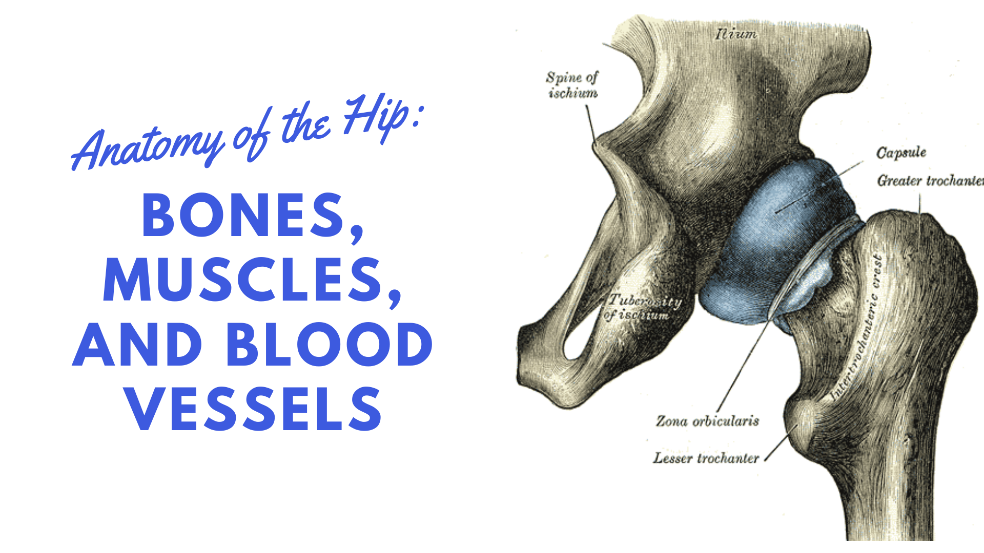
These landmarks are the anterior superior iliac spine.
Human muscle system the muscles of the. The hip itself is a ball and socket joint, much like the shoulder. The distal ends of the radius and ulna bones articulate with the hand bones at the junction of the wrist, which is formally known as the carpus. The lower leg extends from the knee to the ankle. The five metatarsals are the long bones that link the tarsal bones to the toes, seen in yellow in the diagram below. The medial, larger bone of the lower leg. The lower leg is made up of two very strong, long bone—the tibia and the fibula. I think this was the case this is a detailed diagram of a horse's hoof. The majority of muscles in the leg are considered long muscles, in that they stretch great distances. Another bone that is part of the lower leg and the knee joint is called the fibula.this is a bone located on the lateral, or outer part, of the lower leg and is more commonly known as the calf bone. The last pair of the ribs, which is at the bottom of the rib, are called floating. The tibia (shin bone) is the medial bone of the leg and is larger than the fibula, with which it is paired (figure 6.52). Related posts of bones leg diagram picture.
You can palpate its anterior border when you run your finger down the anterior aspect of your leg. You'll learn about the muscle. The humerus is the long bone in the. Hip and leg bone diagram : Subscribe to our free newsletters to receive.
Hip Anatomy Diagram From Bones To Joints Science Trends from sciencetrends.com The knee is a strong but flexible hinge joint that uses muscles and ligaments to withstand the torques and strains of powerful leg movements. The temporal bone is one of the thickest. Bones in leg diagram / tendon and ligament injuries in the horse and recovery. This diagram depicts diagram leg bones anatomy. They are numbered from one to five, starting from the medial (inner) side of the foot. The patella (kneecap) is the sesamoid bone in front of the knee. Leg bone diagram / bones of the human leg 17. It was for a movie the whole time!
The proximal portion of the tibia is tibial plateau which acts as a cusp for the knee, the distal portion tapers into the medial malleoli and the concave surface which articulates with the talus at the ankle joint.
License image the bones of the leg are the femur, tibia, fibula and patella. It was for a movie the whole time! Its lower end helps create the knee joint. Cancellous bone produces red blood cells, platelets, and white blood cells. As these muscles contract and relax they move skeletal bones to create movement of the body. You'll learn about the muscle. Leg bone diagram / bones of the human leg 17. The bone that goes from your pelvis to your knee is called the femur (say: This allows weight to be distributed either anteriorly or posteriorly throughout the foot. The tibia (shin bone) is the medial bone of the leg and is larger than the fibula, with which it is paired (figure 6.52). It is located toward the middle of the lower leg. These landmarks are the anterior superior iliac spine. Also called the shin bone, the tibia is the longer of the two bones in the.